1
/
of
1
Mouse ATG5 (Autophagy Protein 5) ELISA Kit
Mouse ATG5 (Autophagy Protein 5) ELISA Kit
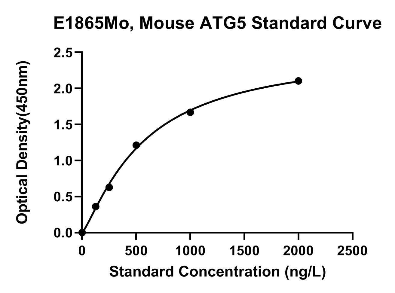
The Mouse (ATG5) Autophagy Protein 5 ELISA Kit measures Autophagy Protein 5 in Mouse samples. The plate has been pre-coated with Mouse ATG5 antibody. ATG5 present in the sample is added and binds to antibodies coated on the wells. And then biotinylated Mouse ATG5 Antibody is added and binds to ATG5 in the sample. Then Streptavidin-HRP is added and binds to the Biotinylated ATG5 antibody. After incubation unbound Streptavidin-HRP is washed away during a washing step. Substrate solution is then added and color develops in proportion to the amount of Mouse ATG5. The reaction is terminated by addition of acidic stop solution and absorbance is measured at 450 nm.
Catalog No:
E1865Mo
Regular price
$595.00 USD
Regular price
$458.00 USD
Sale price
$595.00 USD
Unit price
/
per
2.5 weeks
Couldn't load pickup availability
Product Details
Species Reactivity
Mouse
Sensitivity
9.62 ng/L
Detection Range
20-3500 ng/L
Sample Type
Serum, plasma, cell culture supernates
Incubation(s)
1.5 hour(s)
Research Areas
Cell Biology, Neuroscience, Cancer, Cardiovascular, Metabolism
Background
Involved in autophagic vesicle formation. Conjugation with ATG12, through a ubiquitin-like conjugating system involving ATG7 as an E1-like activating enzyme and ATG10 as an E2-like conjugating enzyme, is essential for its function. The ATG12-ATG5 conjugate acts as an E3-like enzyme which is required for lipidation of ATG8 family proteins and their association to the vesicle membranes. Involved in mitochondrial quality control after oxidative damage, and in subsequent cellular longevity. Plays a critical role in multiple aspects of lymphocyte development and is essential for both B and T lymphocyte survival and proliferation. Required for optimal processing and presentation of antigens for MHC II. Involved in the maintenance of axon morphology and membrane structures, as well as in normal adipocyte differentiation. Promotes primary ciliogenesis through removal of OFD1 from centriolar satellites and degradation of IFT20 via the autophagic pathway. May play an important role in the apoptotic process, possibly within the modified cytoskeleton. Its expression is a relatively late event in the apoptotic process, occurring downstream of caspase activity. Plays a crucial role in IFN-gamma-induced autophagic cell death by interacting with FADD (By similarity). (Microbial infection) May act as a proviral factor. In association with ATG12, negatively regulates the innate antiviral immune response by impairing the type I IFN production pathway upon vesicular stomatitis virus (VSV) infection. Source: UniProt Consortium (2025)
Shipping Condition
Shipped on cold gel packs.
Storage Condition and Shelf Life
2-8C
Analyte
Autophagy Protein 5
Regulatory Status
For Research Use Only